![[N. crassas
asexual life cycle]](asex_gen_cycle.jpg)
Mycel and spore formation in the asexual life cycle of Neurospora crassa
Neurospora crassa has a sexual and an asexual life cycle.
It is during the asexual cycle the sporulation rhythm can be observed.
The picture below indicates the different stages during the asexual
cycle.
Let's begin with the spores: they begin to germinate and form
a mycel at high nutrient conditions and light. The mycel develops
further by forming aerial hyphae which finally bear again conidia.The
pictures below illustrate the various stages in the development
of Neurospora.
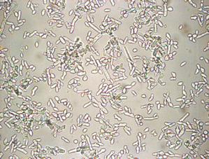
|
Conidia seen in microscope with 400x magnification |
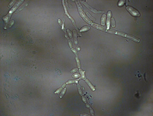
Hyphae and germinating conidia (1000x magnification) |
|
Mycel (100x magnification) |
|
The sporulation rhythm of Neurospora crassa
If a piece of mycel is transferred to a low nutrient medium
and continuous darkness (DD-condition; DD: from German, Dauer-Dunkel,
constant darkness) the circadian oscillator starts.
The oscillations can be seen as regular-repeating zones of sporulation
when the mycel is placed on agar-medium under DD conditions: during
the growth sporulation occurs every 22 h (when using the wild-type
bd-mutant).
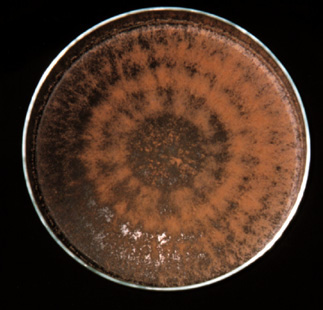
| Rhythmic sporulation of N. crassa observed in a 20 cm petri-dish. A piece of mycel was placed in the middle of the dish which contained agar with minimal medium. After several days in darkness the rhythmic formation of conidia was observed. The color of the conidia is orange due to carotenoids. |
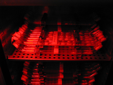
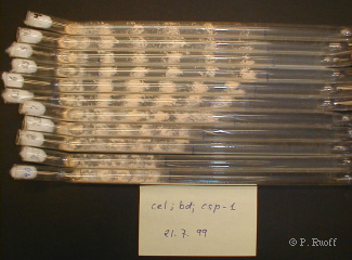
Study of the sporulation rhythm in growthtubes
Normally the rhythm is studied by using petri-plates or so-called
"racetubes". Racetubes are glass tubes that contain
the agar medium.
![[schematic picture of
racetube experiment]](racetube.jpg) |
 |
Racetubes have the advantage that the mycel can grow over a larger
distance and that the period of the sporulation can be easily
determined as the distance between conidiation peaks when the
growth velocity is known. A schematic sideview of a racetube is
shown in the picture above (left). It shows part of the tube where
a piece of mycel has been placed on agar. While the tubes are
kept in continuous darkness, rhythmic bands begins to form. Neurospora
does not respond to red light and work that should be performed
under dark conditions can be done under a red safety light (right
picture above).
|
The photograph to the right shows racetubes seen from above. The growing mycel has formed rhythmic bands of aerial hyphae with spores. The white cotton plugs avoid contamination of the sterile agar medium by other microorganisms while air can still enter the tubes. As ourselves, Neurospora needs oxygen to live! In this particular experiment the extracellular pH in the agar was varied from pH 4 (top racetube) to pH 9 (bottom) with intervals of 0.5 units. It is clearly seen that the growth becomes slower at higher pH. The growth rate is determined by marking the growing front of the mycel every 24 hours. In most instances the growing velocity is on average constant. By determining the growth rate it is then possible to measure the period of the rhythm. This is simply done by measuring the distance between the centers of neighboring sporulation areas and dividing this distance by the growth velocity. Although the growing velocity is depended on pH (and other environmental factors, as temperature, nutrition supply, etc.), the period is generally (and also in this example) found to be unaltered, i.e., around 21.5 hours. This constancy of the period even under different environmental influences (temperature, pH, nutrition) is due to one or several compensation mechanisms within the cells, which keep the period constant. It is now generally believed that circadian rhythms act as time-measuring devices: physiological clocks. |
|
A video of the sporulation rhythm
|
We have extracted two QuickTime® video clips (one 1.9
Mb, the other 3.6 Mb) from a movie taken by Professor emeritus
Van Gooch, University of Minnesota. It shows the rhythmic
sporulation under a red savety light (N. crassa is blind
for red light). Note that in the video the racetubes are seen
from above. click here for the 1.9 Mb video or here for the 3.6 Mb clip |
|
References
Jason C. Thoen and Van Gooch: Time Lapse Video Showing an Internal Circadian Clock
in Mold (Neurospora) Growth.
Rowland H. Davis: Neurospora. Contributions of a Model Organism. Oxford University Press, Oxford, 2000. ISBN 0-19-512236-4.
David D. Perkins, Alan Radford and Matthew S. Sachs: The Neurospora Compendium. Chromosomal Loci. Academic Press, San Diego, 2001. ISBN 0-12-550751-8.
Rowland H. Davis and David. D. Perkins: Neurospora: a model of model microbes. Nature Reviews | Genetics, 2002, 3 , 7-13.

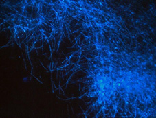
![[picture from sporulation
movie]](Neuro1.gif)