This project is a collaboration between the Department of Pathology at Stavanger university hospital (SUH) and BMDLab at UiS.
Globally there has been an enormous increase in bladder cancer incidents the past decades, with the number of deaths increased by 49% from 1990 to 2010. Correct prognosis of recurrence and progression is essential to avoid under- or over-treatment of the patient, as well as unnecessary suffering and cost. To diagnose the cancer grade and stage, pathologists study the histological images, however, this is a timeconsuming process and reproducibility among pathologists are low.
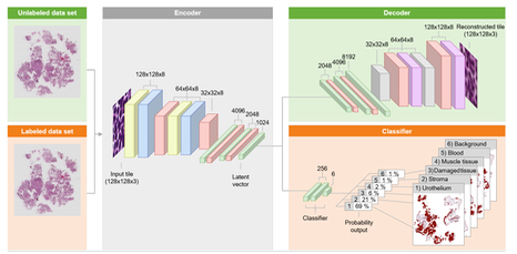
In this project, a method for automatic classification of H&E stained whole slide images (WSI) into six different classes is proposed, where the classes are dened as; urothelium, stroma, muscle tissue, damaged tissue, blood and background. The method is based on convolutional neural networks (CNN), firstly trained unsupervised using large unlabelled image sets by utilizing an autoencoder. A smaller set of labelled images are used to train the final fully connected layers from the low dimensional latent vector of the autoencoder, providing an output as a probability score for each of the six classes, suitable for automatically dening regions of interests in WSI